Uterus and uterine (fallopian) tubes
Label the diagrams
6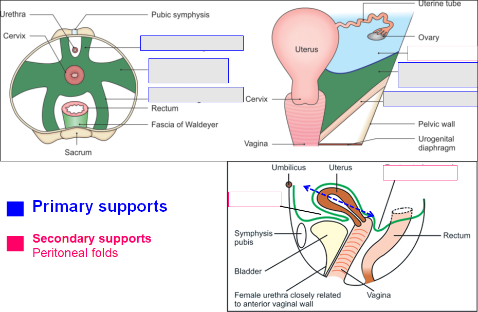
Lable supports of uterus
Viva questions
31What are the parts of uterus?
2What are normal angulations of uterus? What is their significance?
3What are the parts (subdivisions) of uterine (fallopian) tube?
Short-answer questions
8Write note on supports of uterus.
Prolapse of Uterus is prevented (sagging into the vagina) because of the supports of uterus.
Some supports are really significant. They are
PRIMARY supports. -
- Muscular
- Fibromuscular (ligamentous and Mechanical)
SECONDARY supports are not helping very significantly - they are peritoneal folds related to uterus.
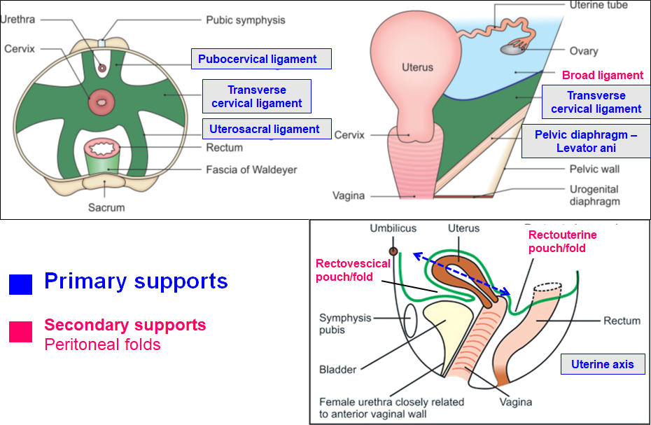
Supports of uterus © BD Chaurasia Vol II
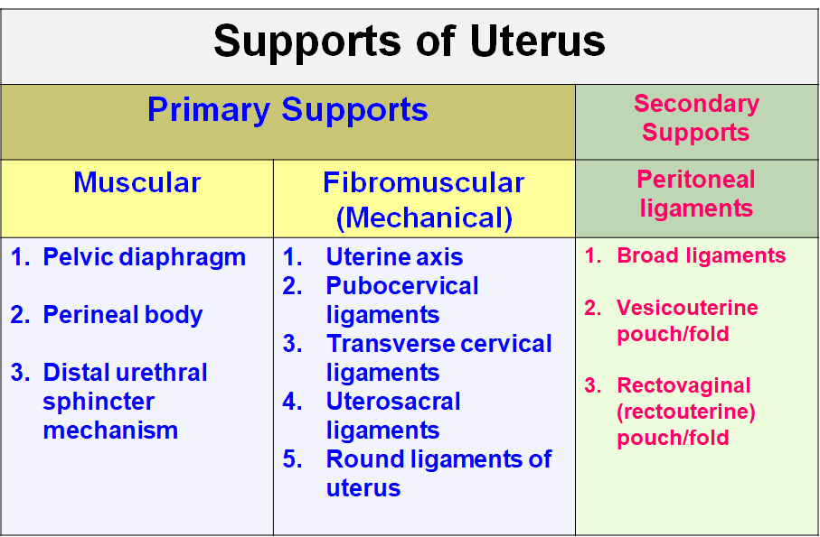
Supports of uterus
Muscular supports
1. Pelvic diaphragm
•Supports pelvic viscera
•Resists increased intra-abdominal pressure
•Fibres of the diaphragm i.e. Pubococcygeus (Pubovaginalis) – form supporting sling around vagina
2. Perineal body
Fibromuscular node – perineal muscles and pelvic diaphragm are attached to it - so maintains integrity of pelvic floor
3. Distal urethral sphincter mechanism
The muscles of urethra - sphincter urethrovaginalis and compressor urethrae are inserted into vagina, thus support vagina and uterus
Fibromuscular supports
1.Uterine axis
Sagging of uterus into vagina is prevented by anteverted and anteflexed position of uterus
*Round ligament pulls fundus of the uterus forwards while the uterosacral ligaments pull the cervix backwards - thus they help to maintain the anteflexed and anteverted position of uterus.
2.Round ligament of uterus
Fibromuscular bands extending from lateral angle of uterus, come forward, pass through inguinal canal and connect with the labia majora
3.Ligaments (derived from endopelvic fascia)
•Transverse cervical ligaments of Mackenrodt (Cardinal ligaments) – Connect cervix to the lateral wall of pelvis
•Pubocervical ligaments – Connects cervix to inner surface of pubic bone
•Uterosacral ligaments – Connect cervix to 2nd and 3rd sacral vertebrae
Secondary supports
Broad ligaments, Vesicouterine pouch/fold, rectovaginal pouch/fold
Write a note on broad ligament of uterus.
It is a 2 layered fold of peritoneum extending from lateral borders of uterus to the lateral wall of pelvis on both sides.
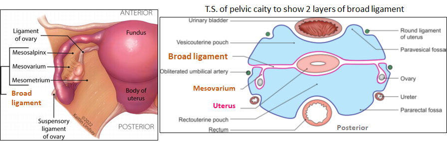
Parts of Broad ligament ©BD Chaurasia Vol II© 2026 Springer Nature
It has
Two surfaces - anterior and posterior
Two borders - upper free & lower attached
Parts -
- Mesosalpinx
- Mesometrium
- Mesovarium
- Suspensory ligament of ovary (infundibulopelvic ligament)
Contents of broad ligament
- one tube - Uterine (Fallopian) tube
- Two ligaments - Round ligament, ligament of uterus
- Two arteries - Uterine artery, ovarian artery
- Two nerve plexuses - Uterovaginal, ovarian
- Three embryonic remanants - Epoophoron & Gartner's duct, Paroophoron, vesciular appendices
- Other - lymph nodes, fibroareolar tissue
Write Clinical applications of uterine (fallopian) tube.
- Tubectomy - for female sterilization. It prevents the meeting of oocyte coming from lateral end and sperm coming from medial end of the tube - thus No fertilisation.
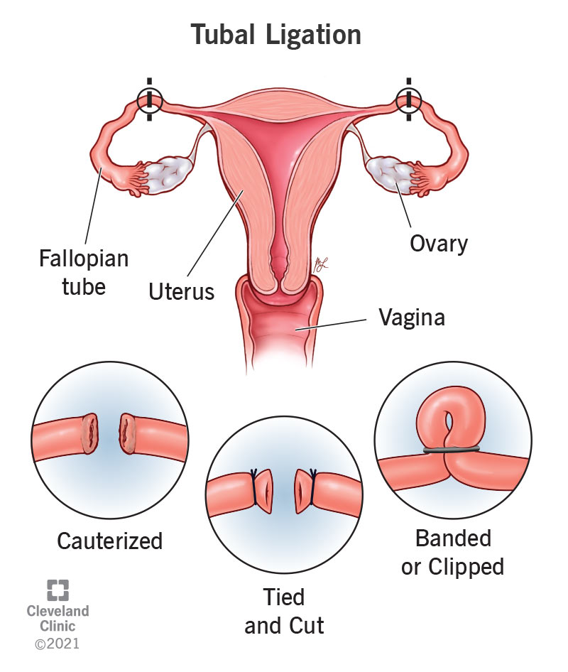
- Sterility due to blockage in fallopian tube - Inflammation of the fallopian tube (salpingitis) is the most common cause of tubal block. Due to the blockage, the male and female gametes cannot come together and fertilization cannot occur.
- Hysterosalpingography - It is a radiograph of uterus and the uteine (fallopian) tubes using contrast medium. A radiopaque dye is injected into the uterine cavity and the tubes. If the tubes are patent, the dye comes out (spillage of dye in peritoneal cavity). No spillage indicates tubal blockage.
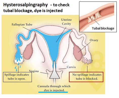
Hysterosalpingography
- Tubal pregnancy - After fertilization, if the embryo gets implanted in the uterine tube - it is tubal pregnancy (ectopic pregnancy). The enlarging embryo can cause rupture of the tube and can lead to life threatening haemorrhage.
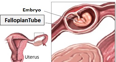
Tubal pregnancy © 1997-2026 ADAM
Write note on microanatomy (histology) of uterus
Wall of the uterus has 3 layers - Innermost Endometrium, middle myometrium (smooth muscles) and outside is perimetrium.
Endometrium
It is lined by simple columnar epithelium.
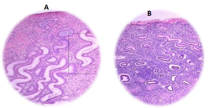
It has 2 layers - Stratum Basalis and stratum functionalis (stratum compactum + stratum spongiosum). Thickness of stratum functionalis goes on increasing during proliferative and secretory phases of uterus. Histologically, uterus looks different in proliferative phase and secretory phase.
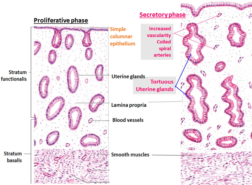
Proliferative vs Secretory phases of uterus© IB Singh Textbook of Histology
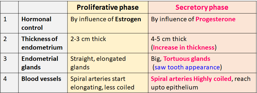
Myometrium - Beneath the stratum basale lies muscular layer of uterus i.e. myometrium. It is thickest layer. Smooth muscle bundles present in this layer are arranged in various directions. Blood vessels supplying the muscle are also seen here.
Write note on development of uterus
No answer yet
Write note on microanatomy of uterine tube.
Wall of the fallopian tube - 3 layers from within outwords - Mucosa, muscular layer, serosa
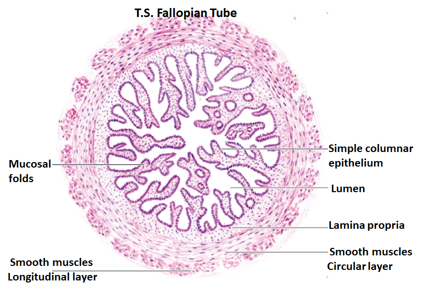
T. S. Fallopian tube © Inderbir Singh Histology
- Mucosa
- It is folded and forms many longitudinal mucosal folds. The folds project into lumen and fill the lumen. The folds are lined by epithelium with underlying lamina propria.
- Epithelium - simple columnar epithelium - with both ciliated and non-ciliated (peg cells) columnar cells
- Non-Ciliated cells (peg cells) - help in capcitation of sperms. Their secretion gives nourishment to sperms and newly formed embryo
- Muscular layer
- Smooth muscles are arranged underneath the mucosa - as inner circular and outer longitudinal layers
- Serosa
- Outer layer of connective tissue and mesothelium
Write clinical anatomy of uterus.
No answer yet
Write a note on uterine artery.
No answer yet
Fill in the blanks
8A forward angle between long axis of body of uterus and long axis of vagina is called ------- and it measures ------- degrees.
A forward angle between long axis of body of uterus and long axis of cervix is called ------- and it measures ------- degrees.
Peritoneal pouch behind the uterus is called -----------.
To repair prolapse of uterus, surgical tightening of ---------- ligaments is most commonly done.
---------- ducts help in development of uterus and fallopian tubes.
Endometrium is lined by ------------ epithelium.
Saw tooth appearance of uterine glands is the characteristic feature of ------------- phase of endometrium.
Image A is ------- phase and image B is ------ phase of endometrium.