scalp & Face
Label the diagrams
3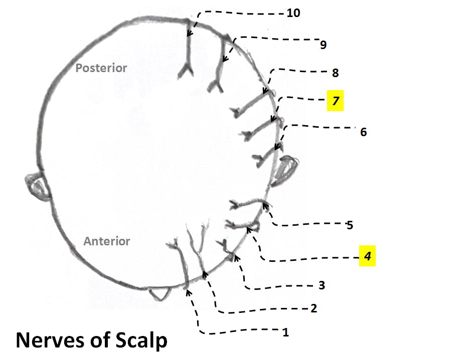
Short-answer questions
4Describe layers of scalp.
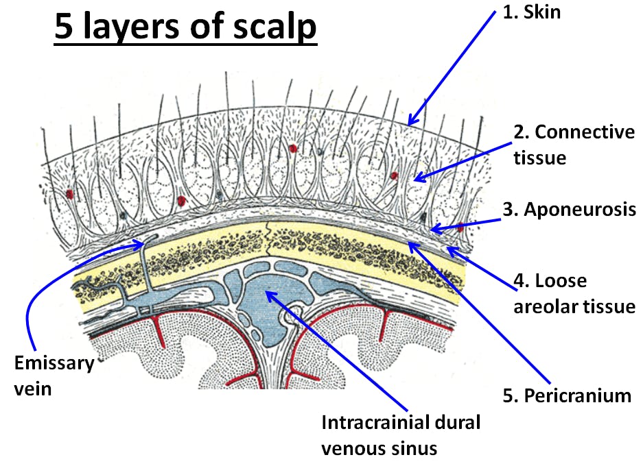
The scalp consists of 5 layers - Skin, Connective tissue, Aponeurosis of occipitofrontalis muscle,Loose areolar tissue, and Pericranium
1. Skin - It is thick, and has numerous hairs, sebaceous and sweat glands
(* It is a common site for the formation of sebaceous cysts)
2. Connective tissue - It is dense subcutaneous fibrous tissue, connects firmly to skin and aponeurois. It consists of blood vessles and nerves of scalp.
(* Walls of blood vessels are adherent to firbrous network of the tissue so in an open wound the vessels get torn and are unable to retract leading to profuse bleeding)
3. Aponeurosis of occipito-frontalis muscle - It a sheet of fibrous tissue connecting frontalis and occipitalis muscles.
(* The fibres of aponeurosis run in antero-posterior direction so the wounds of scalp gape only when the aponeurosis is divided/cut transversly)
4. Loose areolar tissue - it is a potential space beneath the aponeurosis, contains emissary veins
(* The emissary veins connect veins of scalp with intracranial dural venous sinuses. The veins are devoid of valves, so infection of scalp can be transmitted readily into intracrainal sinuses- so the 4th layer is called dangerous area of scalp)
5. Pericranium - is outer periosteum of skull bones - loosely covers the bones except at sutural lines
Describe in short facial artery
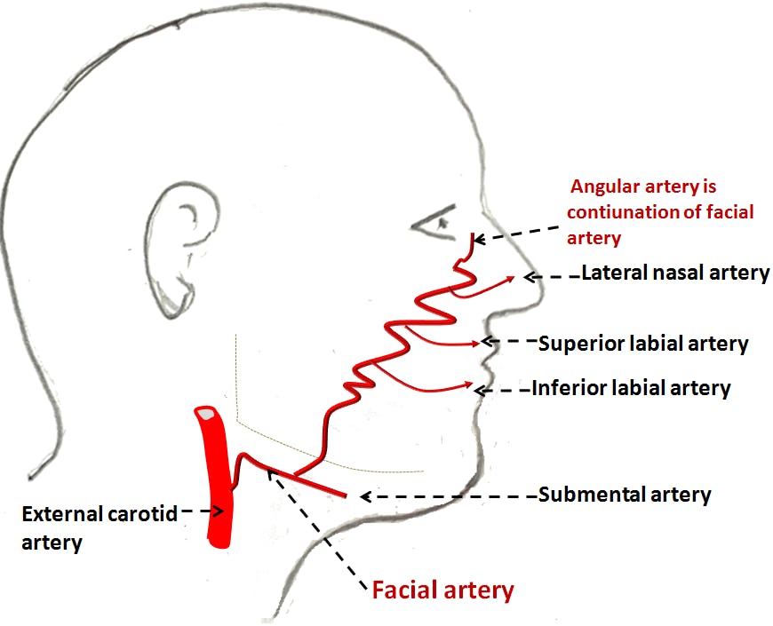
Origin: Facial artery arises from the external carotid artery in the carotid triangle of neck
Course and relations: In the neck the artery is intimately related to submandibular salivary gland. It enters the face by winding round the base of mandible at the anterior border of masseter muscle. Here it lies deep to marginal mandibular branch of facial nerve and anterior to facial vein.
In the face - its course is tortuous. It passes upwards and forwards lateral to angle of mouth and then ascends along the side of the nose
Branches:
In the neck it gives - ascending palatine, tonsillar, submental branches and branches to submandibular gland
In the face it gives - Inferior labial, superior labial and lateral nasal branches
* Anastomoses
Anastomoses with transverse facial artery
Anastomoses between right and left superior labial and right and left inferior labial arteries (**hence wounds of lip bleed freely)
Distal to lateral nasal branch it continues as angular artery and anastomoses with a branch of ophthalmic artery (** thus a free communication between internal and external carotid arteries)
Describe nerve supply of scalp
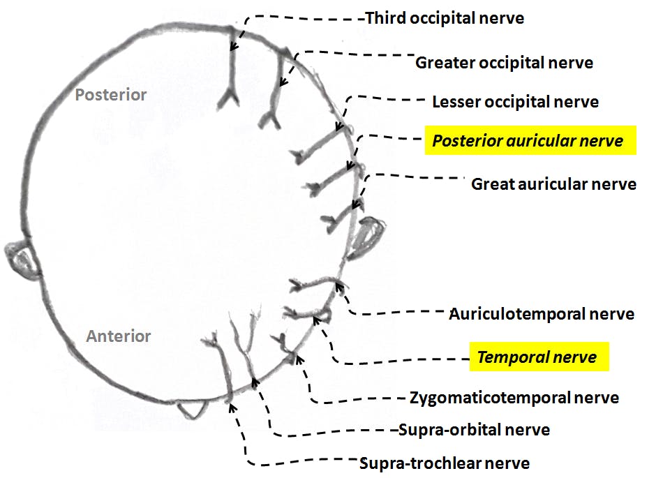
The scalp is supplied by 10 nerves on each side - 5 nerves in front of auricle and 5 nerves behind. In each set of 5 nerves there is 1 motor nerve and 4 sensory nerves.
Nerves in front of auricle from before backwards -
1. Supra-trochlear nerve(sensory)
2. Supra-orbital nerve (sensory)
3. Zygomatico-temporal nerve(sensory)
4. Temoporal branch of facial nerve (*motor) to frontalis muscle
5. Auriculo-temporal nerve (sensory)
Nerves behind the auricle from before backwards:
6. Great auricular nerve (sensory)
7. Posterior auricular branch of facial nerve (*motor) - to occipitalis muscle
8. Lesser occipital nerve (sensory)
9. Greater occipital nerve (sensory)
10. Third occipital nerve (sensory)
Describe the blood supply of scalp
Scalp is rich in blood supply. (So wounds of scalp bleed profusely , but they heal faster)
Arterial supply
It is supplied by 5 arteries on each side (3 in front of auricle and 2 behind)
1. Supra-trochlear artery
2. Supra-orbital artery
3. Superficial temporal artery
4. Posterior auricular artery
5. Occipital artery
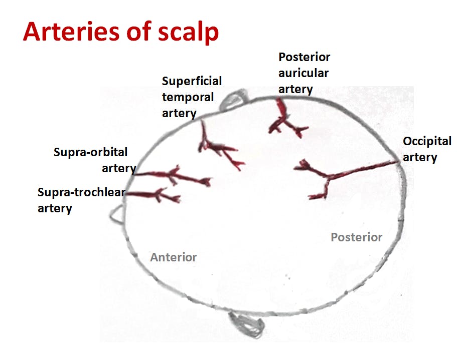
* First 2 arteries are indirect branches of Internal carotid artery and remaining 3 arteries are branches of external carotid artery. These 5 arteries freely anastomose with each other. So scalp is a site of anastomosis between Internal and External carotid arteries.
Venous drainage
Veins of scalp correspond to the arteris.