Cranial meninges & dural venous sinuses
Label the diagrams
1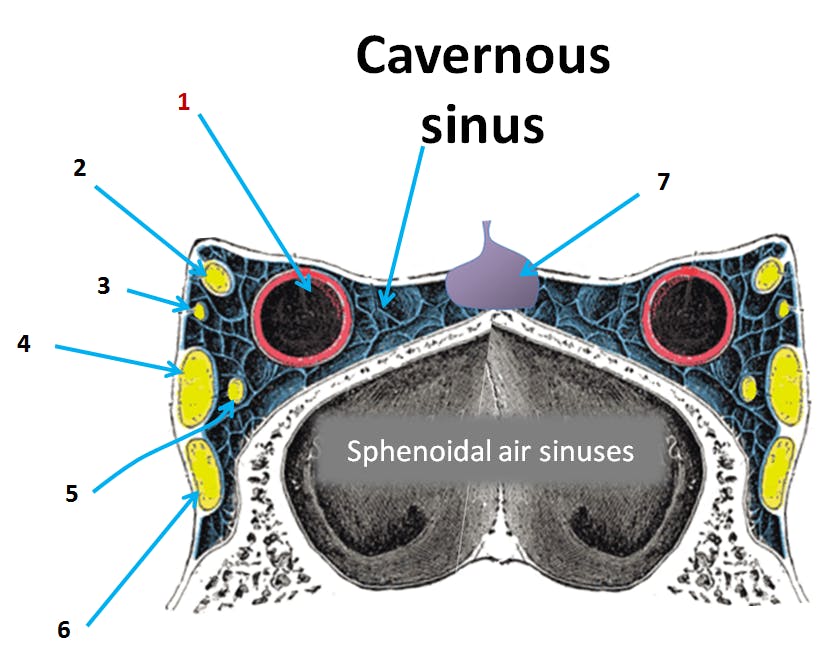
Viva questions
31Name any 4 unpaired dural venous sinuses
2Name any 4 paired venous sinuses
3Enumerate any 4 tributaries of cavernous sinus
Short-answer questions
3Describe location of cavernous sinus and the structures passing through it with its clinical anatomy
Location: Cavernous sinuses are a pair of dural venous sinuses, located in the middle cranial fossa on either side of the body of sphenoid.
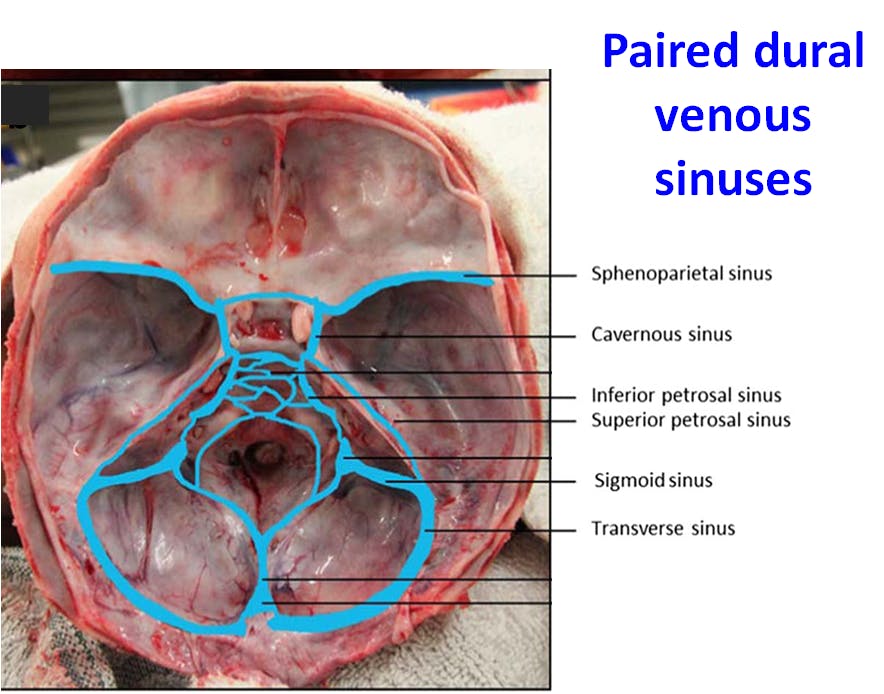
By Emma C. Cheshire, Roger D. G. Malcomson, Shiju Joseph, Asif Adnan, David Adlam, Guy N. Rutty - Cheshire, E.C., Malcomson, R.D.G., Joseph, S. et al. Int J Legal Med (2017) 131: 1377. https://doi.org/10.1007/s00414-017-1570-1, CC BY 4.0, https://commons.wikimedia.org/w/index.php?curid=79731841
Structures passing through cavernous sinus:
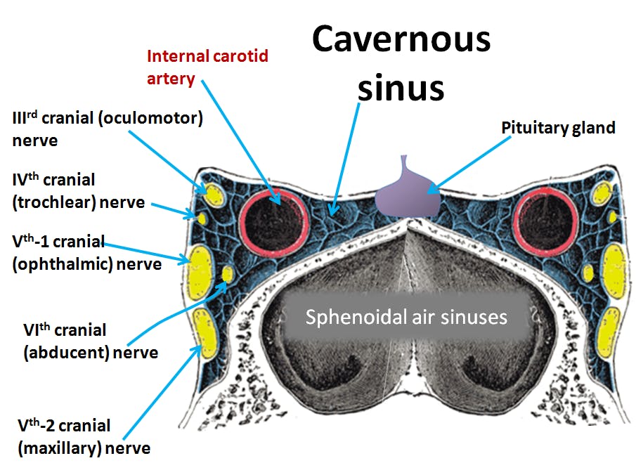
* Structures in the lateral wall of the sinus from above downwards are -
Oculomotor (III), trochlear (IV), ophthalmic division of trigeminal (V) and maxillary division of trigeminal (V) nerves
* Structures near the floor of the sinus are -
Internal carotid artery and abducent (VI) nerve
Clinical Anatomy
1. Pulsating Exophthalmos: During fracture of skull, internal carotid artery is ruptured - and arteriovenous communication is formed between the artery and cavernous sinus. The eyeball is protruded (exophthalmos) and pulsates with the arterial pulse. Venous congestion causes edema of eyelids
2. Cavernous sinus thrombosis can lead to injury to the nerves passing through it - leading to paralysis of extraocular muscles and ptosis (III, IV, VI nerves) and pain in eye and forehead (ophthalmic division of V)
Describe communications of cavernous sinus with its clinical anatomy
Communications of cavernous sinus
1. With transverse sinus - via superior petrosal sinus
2. With internal jugular vein - via inferior petrosal sinus
3. With pterygoid venous plexus - through emissary veins
4. With facial vein - by 2 routes
- via superior ophthalmic and angular veins
- via pterygoid venous plexus and deep facial vein
5. With opposite cavernous sinus - via anterior and posterior intercavernous sinuses
6. With superior sagittal sinus - via superficial middle cerebral vein
Clinical Anatomy
Cavernous sinus thrombosis: can be caused due to infection spread via the communications e.g. via dangerous area of face. Thrombosis can lead to -
- Exophthalmos and edema of eyelids due to engorgement of ophthalmic veins
- paralysis of extraocular muscles due to involvement of III, IV and VI cranial nerves
- Pain in the eye and forehead due to involvement of ophthalmic division of trigeminal nerve
- Ptosis due to injury to III cranial nerve
Write a note on superior sagittal sinus
Superior sagittal sinus is one of the unpaired dural venous sinuses.
Location: It lies within the covex attanched margin of the falx cerebri.
Course: The sinus begins at the crista galli. As it passes backwards it increases in size. When it reaches posteriorly at internal occipital protuberance, it deviates to the right and becomes continuous with right transverse sinus. Confluence of sinuses is the dilated posterior end of superior sagittal sinus.
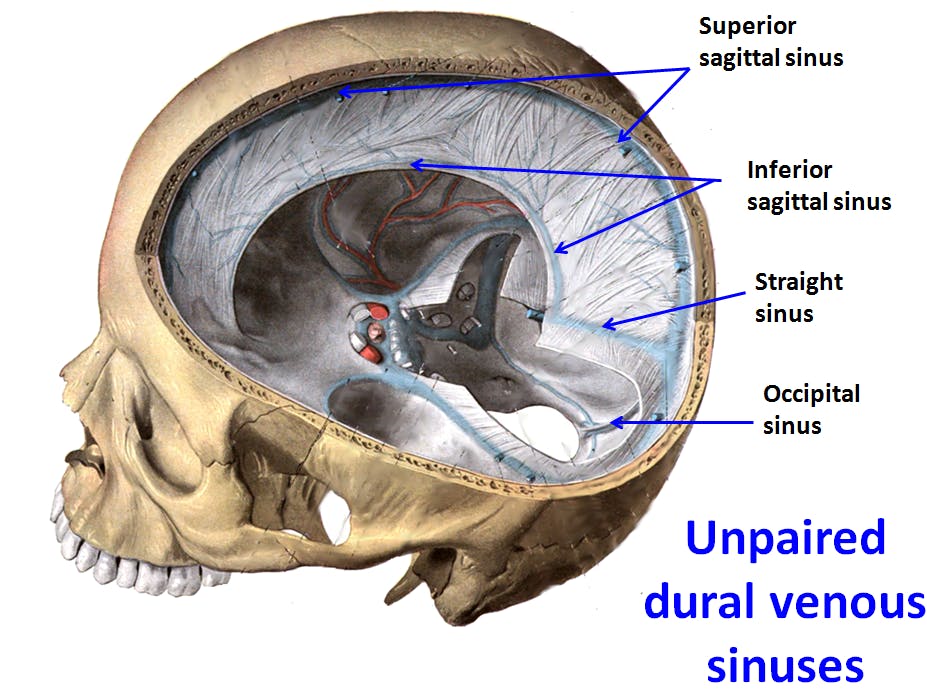
Public Domain
Tributaries: About 8-12 Superior cerebral veins and diploic and emissary veins.
* Communications:
- with veins of scalp through parietal emissary veins
- with cavernous sinus through superficial middle cerebral vein
Interior of the superior sagittal sinus shows :
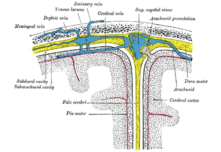
- Opening of superior cerebral veins
- Arachnoid granulations projecting into it (CSF present in it is absorbed into the sinus)